R & F® Listeria sp./L. monocytogenes Chromogenic Plating Medium

R & F® Listeria sp./L. monocytogenes Chromogenic Plating Medium

Request a Quote
Selective/Differential Medium For Detecting Nonpathogenic & Pathogenic Listeria On A Single Plate

Colony Appearance

Presumptive positive colonies of nonpathogenic Listeria sp. appear pink, whereas Listeria monocytogenes (L.m.) and Listeria ivanovii (L.i.) appear as blue-green to blue-violet colonies after 42-48 h at 35°C. A small percent of Enterococcus sp. will grow as white colored colonies. Bacillus sp., yeasts and Gram (-)s are inhibited. A quick and simple biochemical test on our confirmation medium biplate can easily distinguish, within 6 h, L. m. (fluorogenic reaction and/or acid production) from L.i. (negative for both).

Mechanism

Our Listeria sp./Listeria monocytogenes medium, as in the Listeria monocytogenes medium, contains the chromogenic substrate X-inositol phosphate (X-IP) that detects the enzyme phosphatidylinositol phospholipase C, virulence factor, which is found in L.m. and L.i. producing blue-green or blue-violet colonies. Even though L.m. and L.i. are the same color, L.i. strains are very seldom found in foods. In addition, this medium will also detect other Listeria sp. by using the combination chromogenic substrates of Salmon-β-D-glucopyranoside and Magenta-β-D-glucopyranoside (detects the β-glucosidase enzyme) in which these strains will produce pink colonies against an opaque white background (titanium oxide). Our confirmatory medium biplate uses a quick fluorogenic reaction in α-mannosidase and an acid production from rhamnose to distinguish L.m. from L.i. 

Industry Types

Fish, poultry, meat, dairy, bakery and ready-to-eat foods and environmental testing in food processing facilities.

See Product Brochure, Use Protocol, SDS, Certificate of Analysis and Medium Preparation for more information about our medium.

Advantages/Benefits

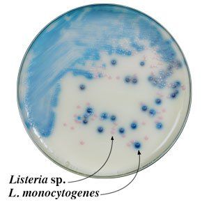
1. Highly differential medium that works by the combination of indoxyl derivative chromogenic substrates that produce positive color reactions for colonies of nonpathogenic Listeria sp. that are pink due to their ß-glucosidase activity, and blue-green to blue-violet for the pathogenic species depending on the strain-specific balance of ß-glucosidase (pink) and phosphatidylinositol-specificphospholipase C (blue) activities.

2. Other companies’ selective media for Listeria species that only depend on the detection of ß-glucosidase activity produce a single color for colonies for all Listeria species.

3. The agar surface of our plates have an opaque white background that facilitates differentiation of colored colonies of both groups of Listeria organisms growing on the surface of the plate.

4. Our medium’s selectivity and sensitivity for the pathogenic Listeria species (L.m. & L.i.) is 96% and 100%, respectively, with no false negatives.

5. Water insoluble chromogenic lattice prevents the colony color from diffusing into the agar making detection and enumeration much easier.

6. Competitors’ non-chromogenic agars (MOX, Oxoid & Palcam) which depend on esculinase as the differential trait allows many Listeria sp., Bacillus sp. and Enterococcus sp. to grow as black color colonies. Since the end product is water soluble, the black color will diffuse into the medium. Additional expensive tests are required to further identify L.m. strains.

7. Competitors’ chromogenic media which utilize the X-β-D-glucopyranoside substrate will allow too many Listeria sp., Bacillus sp. and Enterococcus sp. to grow as grow as blue colored colonies without clear halos. These colonies will interfere with the identification of L.m. & L.i. by coalescing together making it difficult to see the clear halo around the blue colony of the L.m. & L.i.

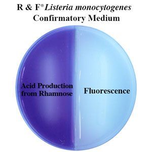
8. Our rapid and convenient fluorogenic/acid test differentiates L.m. from L.i. as quick as 6 hours-saving time and costs.

9. The shelf life of prepared plates remains stable for at least 60 days, stored in the dark at 2-8°C. One bottle of plating medium powder and two boxes of supplements will make approximately 230 plates.

Publications

1. Restaino, L., E.W. Frampton, W.C. Lionberg, and A.L. Restaino. 2006. A multi-chromogenic agar for the dual detection of nonpathogenic and pathogenic Listeria species. Poster No. P3-64, IAFP 2006.

2. Restaino, L., E.W. Frampton, W.C. Lionberg, and A.L. Restaino. 2007. Detection of non pathogenic and pathogenic Listeria species by use of a chromogenic agar. Food Prot. Trends. 27: 592-596.

3. Swiech, R. et al. 2008. Efficacy of a chromogenic plating medium for detaching Listeria species from environmental samples. Poster No. P3-16, IAFP 2008.

Ordering Information

M-1150
R & F® Listeria sp./Listeria monocytogenes Chromogenic Detection System (250 g Bottle of Powder & two Supplement Boxes – Makes 220-240 Plates)
M-1100
R & F® Listeria sp./Listeria monocytogenes Chromogenic Plating Medium (250 g Bottle)
M-1110
R & F® Listeria sp./Listeria moncytogenes Supplement for Plating Medium (1 Box)
M-0520
R & F® Listeria moncytogenes Confirmatory Medium (100 g Bottle)
M-1160
R & F® Listeria sp./Listeria monocytogenes Chromogenic Prepared Plate (Minimum Order of 20 Plates – Shelf life is 60 Days)
M-0570
R & F® Listeria monocytogenes Confirmatory Prepared Biplate (Minimum Order of 20 Plates – Shelf life is 60 Days)
Share by: